Home

mydlo anjel nahrávať time lapse microscopy kalórie urýchľovač myšlienka

Current Status of Time-Lapse Microscopy for Embryo Selection | SpringerLink

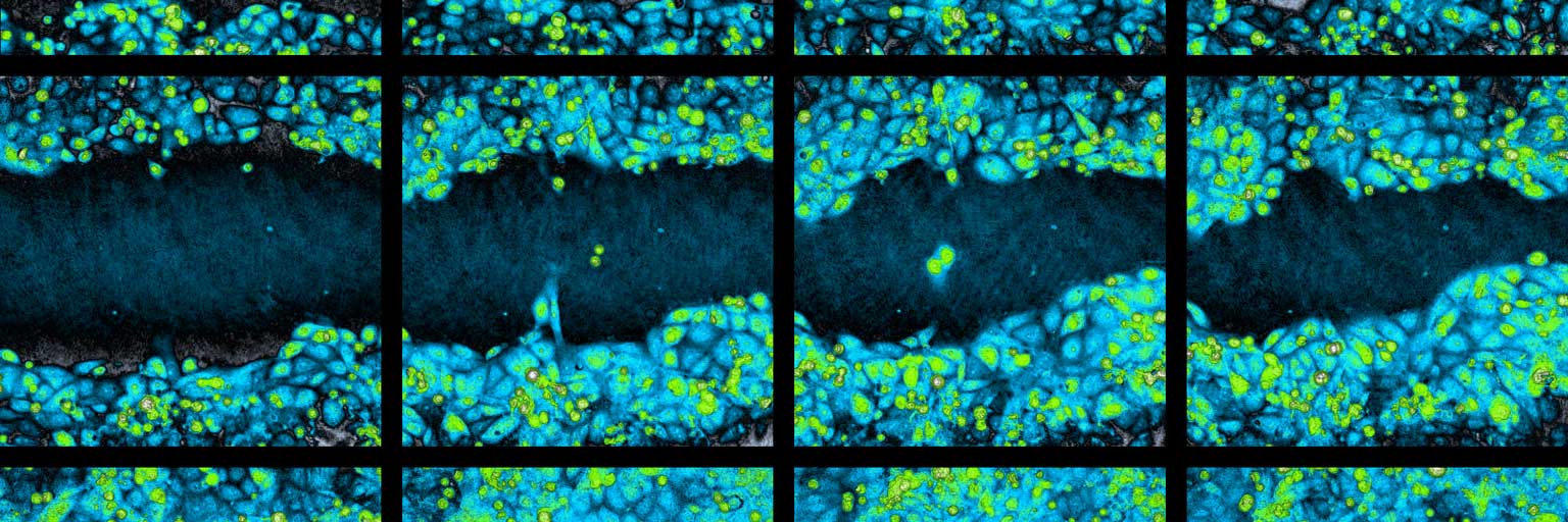
Time-Lapse Microscopy Captures Cells in the Act | Features | Apr 2011 |  BioPhotonics
Time-Lapse Microscopy Captures Cells in the Act | Features | Apr 2011 | BioPhotonics

New image analysis method for time-lapse microscopy shows how giant viruses  infect amoeba | Editage Insights
New image analysis method for time-lapse microscopy shows how giant viruses infect amoeba | Editage Insights

Through the Looking Glass: Time-lapse Microscopy and Longitudinal Tracking  of Single Cells to Study Anti-cancer Therapeutics | Protocol
Through the Looking Glass: Time-lapse Microscopy and Longitudinal Tracking of Single Cells to Study Anti-cancer Therapeutics | Protocol
Imaging Cytometry by Holographic Time-lapse Microscopy - PHI

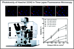
Phototoxicity of Hoechst 33342 in time-lapse fluorescence microscopy -  Photochemical & Photobiological Sciences (RSC Publishing)
Phototoxicity of Hoechst 33342 in time-lapse fluorescence microscopy - Photochemical & Photobiological Sciences (RSC Publishing)

Time-Lapse Microscopy - an overview | ScienceDirect Topics

Applied Sciences | Free Full-Text | ACDC: Automated Cell Detection and  Counting for Time-Lapse Fluorescence Microscopy
Applied Sciences | Free Full-Text | ACDC: Automated Cell Detection and Counting for Time-Lapse Fluorescence Microscopy

A novel apparatus for time-lapse optical microscopy of gelatinisation and  digestion of starch inside plant cells - ScienceDirect
A novel apparatus for time-lapse optical microscopy of gelatinisation and digestion of starch inside plant cells - ScienceDirect

Motion Compensation in Time-Lapse Microscopy - YouTube

New image analysis method for time-lapse microscopy shows how giant viruses  infect amoeba
New image analysis method for time-lapse microscopy shows how giant viruses infect amoeba

Live Cell Imaging | Biomedical Imaging Unit | University of Southampton

Items - Queen Mary University of London

Time-lapse Microscopy | University of Tübingen

Monitoring bacterial growth by time-lapse microscopy. (A) Phase... |  Download Scientific Diagram
Monitoring bacterial growth by time-lapse microscopy. (A) Phase... | Download Scientific Diagram

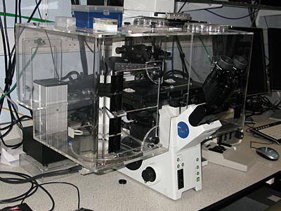
Extended Time-Lapse Microscope

Time lapse microscopy analysis of MV infectious-center formation in... |  Download Scientific Diagram
Time lapse microscopy analysis of MV infectious-center formation in... | Download Scientific Diagram

Time-lapse microscopy images of A2780CP70 cells using a confocal... |  Download Scientific Diagram
Time-lapse microscopy images of A2780CP70 cells using a confocal... | Download Scientific Diagram

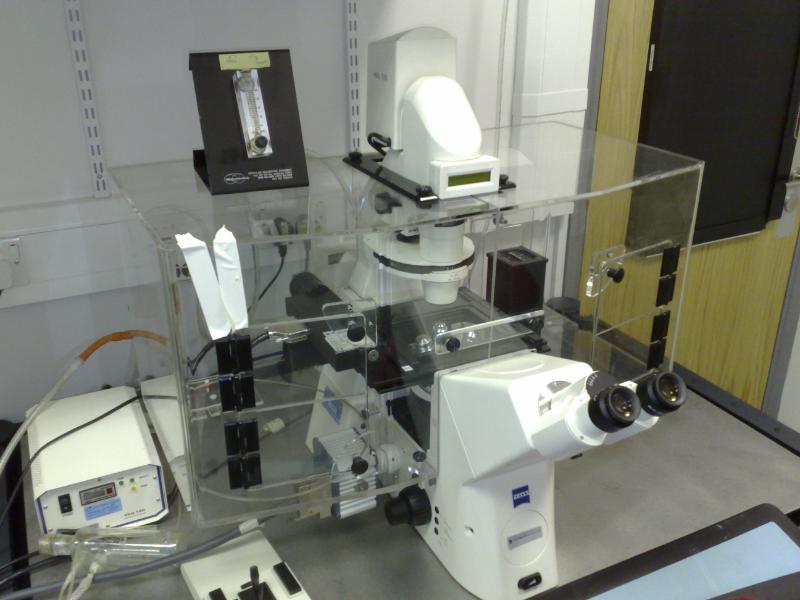
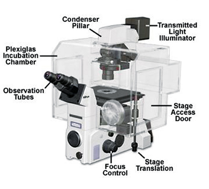
Environmental Considerations for Long Term Timelapse Imaging - 2008 - Wiley  Analytical Science
Environmental Considerations for Long Term Timelapse Imaging - 2008 - Wiley Analytical Science

Cell Biology imaging index

Time-lapse microscopy - Wikipedia

August 4, 2020 | Spencer Lab | University of Colorado Boulder

A Modular and Affordable Time-Lapse Imaging and Incubation System Based on  3D-Printed Parts, a Smartphone, and Off-The-Shelf Electronics
A Modular and Affordable Time-Lapse Imaging and Incubation System Based on 3D-Printed Parts, a Smartphone, and Off-The-Shelf Electronics

Time-lapse confocal imaging datasets to assess structural and dynamic  properties of subcellular nanostructures | Scientific Data
Time-lapse confocal imaging datasets to assess structural and dynamic properties of subcellular nanostructures | Scientific Data